. 2021 Sep 6;10:e69377. doi: 10.7554/eLife.69377

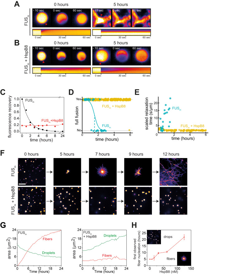
Figure 3. HspB8 prevents hardening and fiber formation of FUS droplets and keeps them dynamic.

(A) FRAP experiment of fresh FUSm condensates (0 hr) and condensates incubated for 5 hr. A kymograph shown below illustrates the kinetics of the process. (B) FRAP experiment of fresh FUSm condensates mixed with HspB8 (0 hr) and condensates incubated for 5 hr. A kymograph shown below illustrates the kinetics of the process. (C) Kinetics of the FUSm aging process. Plotted are the initial slopes of the FRAP recovery curves for FUSm condensates in the absence (black) or presence of HspB8 (red). (D) Successful complete fusion events were registered over time, demonstrating the aging process of the FUSm sample in the absence (turquoise, N=40) or the presence of HspB8 (yellow, N=330). The half-life of liquid-like FUSm condensates alone was estimated to be around 1.5 hr from logistic regression. (E) The size-normalized coalescence relaxation time is an indicator for the material state of the condensates. While it increases for FUSm condensates during the hardening process, it stays constant over 6 hr in the presence of HspB8. Tweezer experiments were performed with fresh samples of 5 μM FUSm with or without 20 μM HspB8. (F) Aging process of 5 µM FUSm condensates in the absence and presence of 5 µM HspB8. In the presence of the chaperone, the droplet morphology is maintained over the whole timeframe of the experiment (12 hr). Scale bar is 10 µm. (G) Left panel: shown is the total area of droplet material (green line) or fibrous material (red line) within FUSm droplets as a function of time after FUSm droplets were added to the existing fibrous material. Spacing between data points is 30 min. Right panel: shown is the total area of droplet material (green line) and fibrous material (red line) within FUSm droplets as a function of time after FUSm droplets and HspB8 were added to the existing fibrous material. Spacing between data points is 30 min. (H) The onset of 5 µM FUSm fiber formation as a function of HspB8 concentration. FRAP, fluorescence recovery after photobleaching.

Figure 3.

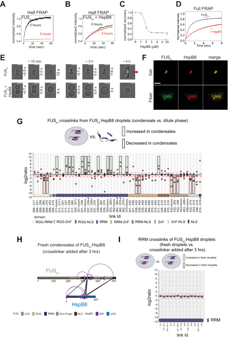
Figure 3—figure supplement 1. HspB8 prevents hardening and fiber formation of FUS droplets and keeps them dynamic.

Figure 3—figure supplement 1.

(A) Quantification of a half-bleach FRAP experiment of fresh FUSm condensates (black) and condensates incubated for 5 hr (red). (B) Same experiment as in (A) in the presence of 5 µM HspB8. (C) HspB8-dependent decrease in mobility has an EC50 of 1.5 µM. Plotted are the initial slopes of the FRAP recovery curves for 5 µM FUSm condensates in the presence of different concentrations of HspB8. (D) Quantification of a full-bleach FRAP experiment of fresh FUSm condensates in the absence (black) and presence of 5 µM HspB8 (red). (E) Representative images of attempted fusion events at early and late stages. Attempts to fuse several droplets of FUSm condensates after hardening result in large amorphous assemblies (red arrow). Scale bar is 3 μm. (F) Colocalization of gels or fibers formed by FUSm and Cy3-labeled HspB8. Scale bar is 10 µm. (G) Crosslink abundance plot from reconstituted FUSm:HspB8 condensates. Plotted are the relative enrichment (droplet vs. non-droplet) for each unique crosslinking site (y-axis) sorted according to the known domain structure within FUSm and HspB8 (x-axis). Shown are only high confidence crosslinking sites (see Materials and methods for details) from five biologically independent sets of experiments (n=5; circles in different shades of gray). Crosslinking sites that were consistently upregulated or downregulated twofold or more (log2ratio≥1 or ≤−1 and FDR≤0.05) in at least two out of five biological replicate sets of experiments and in addition contained no opposing regulation in any replicate set were considered significant and are highlighted with a green (enriched in droplets) or red background rectangle (decreased in droplets). All other changes in crosslinking abundances were considered insignificant and are shown on gray background. The significance threshold of twofold enrichment is indicated as dashed red lines. Dimeric links are indicated by an additional ‘−d’, loop-links by a ‘−l’, and mono-links by an ‘-mono’ at their respective unique crosslinking site. (H) Overall crosslinking pattern of FUSm:HspB8 condensates that were allowed to mature and that were crosslinked 3 hr after their initial formation. (I) Focus on the RRM domain. A comparison of the crosslinks within the RRM (intra, loop, and mono-links) shows no change in abundance when FUSm:HspB8 condensates of fresh droplets were compared with those that were crosslinked 3 hr after their initial formation. FRAP, fluorescence recovery after photobleaching; RRM, RNA recognition motif.