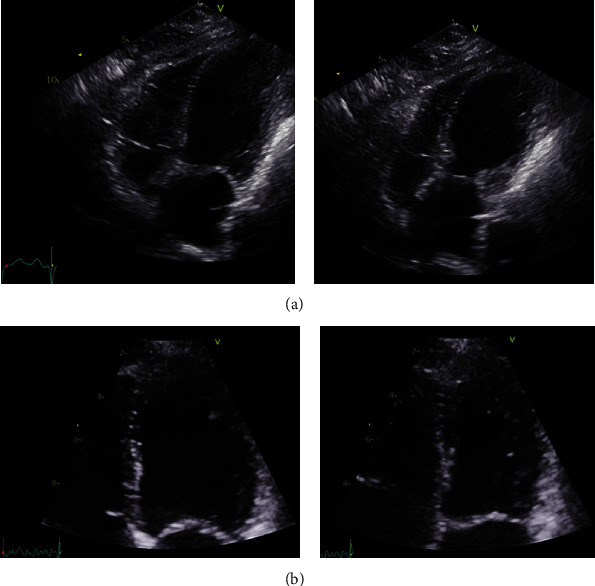
Figure 1.

(a) Echocardiography in the acute phase. The first echocardiography showing modified 4 ch view of end-diastolic and end-systolic left ventricular volume. Basal hypercontraction and apical ballooning is evident, interpreted as acute heart failure/takotsubo cardiomyopathy. (b) Echocardiography after remission of tension pneumothorax and subcutaneous emphysema. Follow-up echocardiography showing 4 ch view of end-diastolic and end-systolic left ventricular volume 1 month later. Left ventricular ejection fraction (LVEF) has normalized.
