Fig. 1.
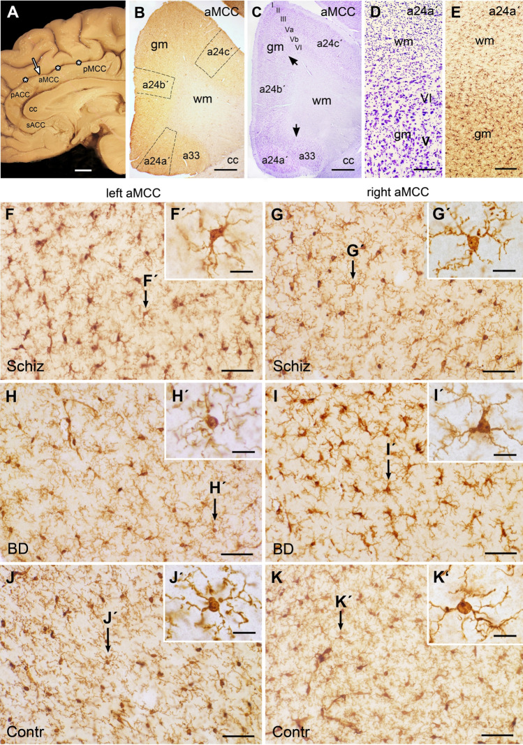
Investigated areas and morphology of microglia in the midcingulate cortex (aMCC). a Medial sagittal surface view shows the corpus callosum (cc), the overlying aMCC between the perigenual anterior cingulate (pACC) and posterior midcingulate cortex (pMCC) and the cingulate sulcus (asterisks). Arrow marks the investigated area. b Overview of the Iba1-stained cryosection shows the aMCC areas a24a′, a24b′ and a24c′ with the marked counted fields. c In the adjacent cresyl violet-stained section the borders between the subareas are marked by black arrows. Note the cortical layers I, II, III, Va, Vb, VI in the grey matter (gm). d Enlargement of the cresyl violet section shows the border area of white matter (wm) with abundant glial cells in contrast to the neurons in the cortical layers V and VI of the gm. e Note the difference in Iba1-staining between wm and gm. In the wm the distribution of microglial cell processes are oriented between the light appearing myelinated nerve fibers; in the gm numerous branching microglial processes extend in all directions from the somata. f–k Micrographs of Iba1-stained examples show the left (f, h, j) and right aMCC (g, i, k) of schizophrenia (Schiz; f, g), bipolar disorder (BD; h, i) and controls (contr; j, k) all of which with ramified microglia with slightly varying phenotypes confirmed by the inserts (f, g′–k′). Bar in a = 1 cm; bar in b and c = 1 mm; bar in d and e = 100 µm, bar in f–k = 50 µm, bar in f′–k′ = 10 µm. sACC subcallosal anterior cingulate cortex

