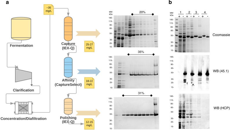
Fig. 3. Production of ProC6C in L. lactis.
a Schematic representation of fermentation and three purification steps of ProC6C protein with associated yields and recovery of purified protein. Coomassie blue-stained 4–12.5% polyacrylamide gel inset with elution fractions (concentration B buffer); upper panel HiPrep Q HP column, middle panel Capture selectXL column, and lower panel HiPrep Q HP. The sample was loaded without a reducing agent. b Analysis of purified ProC6C by SDS-PAGE; upper panel; 1 diafiltrate, 2 HiPrep Q HP column, 3 Capture selectXL column, 4 HiPrep Q HP column purified ProC6C. An immune-blot analysis of the same gel shown in the upper panel using mAb45.1 and anti-L. lactis antibodies (detection of HCP) in the middle and lower panels, respectively. Protein was loaded in each lane with (+) or without (−) DTT (10 mM). The sizes (kDa) of the molecular mass markers are indicated. All blots and gel are derived from the same experiment and processed in parallel.

