
Figure 1:
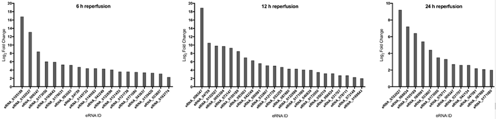
Differentially expressed eRNAs in the post-stroke female cortex at multiple time-points of reperfusion. Expression levels of significantly altered eRNAs determined using qPCR in the post-stroke female cortex at 6, 12 and 24 h of reperfusion, as compared to sham. Values represent average fold-changes determined using the ΔΔCT method from four biological replicates per group, tested in duplicate. X-axis: eRNA ID; Y-axis: Log2 fold-change.