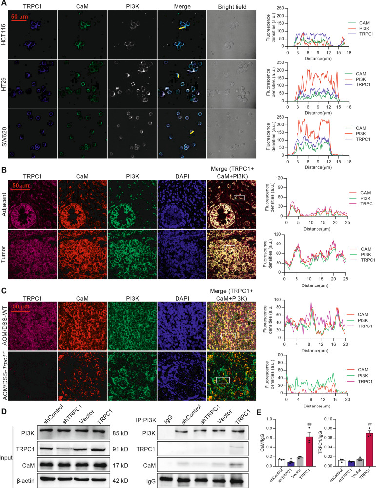
Fig. 6. TRPC1 enhances the interaction between CaM and PI3K in colorectal cancer.
A The colocalization of TRPC1, CaM, and PI3K in CRC cells using immunofluorescence analysis. The representative immunohistological staining of TRPC1, CaM, and PI3K in the HCT116, HT29, and SW620 cell lines (left panel) and the traces of fluorescence intensity spatial profiles (right panel). B The representative immunohistological staining of TRPC1, CaM, and PI3K in human colorectal tumor tissues and adjacent tissues from the local cohort (left panel) and the traces of fluorescence intensity spatial profiles (right panel). Nuclear were stained with DAPI. C Trpc1 knockout inhibited the colocalization of CaM and PI3K. Left panel, the representative immunohistological staining of TRPC1, CaM, and PI3K in CRC tumor tissues in the AOM/DSS-WT and AOM/DSS-Trpc1-/- mice. Right panel, the traces of fluorescence intensity spatial profiles. D, E The interaction of TRPC1, CaM, and PI3K p85 subunit was confirmed by co-immunoprecipitation assay in SW620 cells. Typical results are shown in the left panel along with the statistical analysis in the right panel (n = 3). *P < 0.05 vs. shControl group; ##P < 0.01 vs. vector group.

