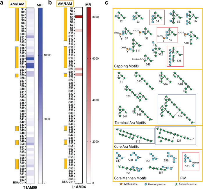
Fig. 3. Reactivity of mAbs T1AM09 and L1AM04 to mycobacterial envelope glycan epitopes.
Median Fluorescence Intensity (MFI) of a T1AM09 (5 µg/mL), and b L1AM04 (5 µg/mL) binding 63 mycobacterial oligosaccharide fragments. Arabinomannan/lipoarabinomannan (AM/LAM) specific fragments (S#1–12, 15–22, 25, 44, 45, 49, 50, 56–59) are marked by the yellow side bar. Six other glycans on the array are: α-glucan (S#13, 14, 24, 46, 48, 52), trehalose mycolates and lipooligosachharides (LOSs; S#38, 39, 54, 55), phenolic glycolipids (PGLs; S#26–37, 40–43, 50, 53), phosphatidyl-myo-inositol mannosides (PIMs, S#23) and glycopepitdolipids (GPLs; S#47, 60, 61). c AM/LAM and PIM motifs with those most strongly recognized by T1AM09 (blue) and L1AM04 (red). Each data point (S1–S63) represents the mean from two or more replicates. All bonds are alpha unless designated in the figure as beta (β).

