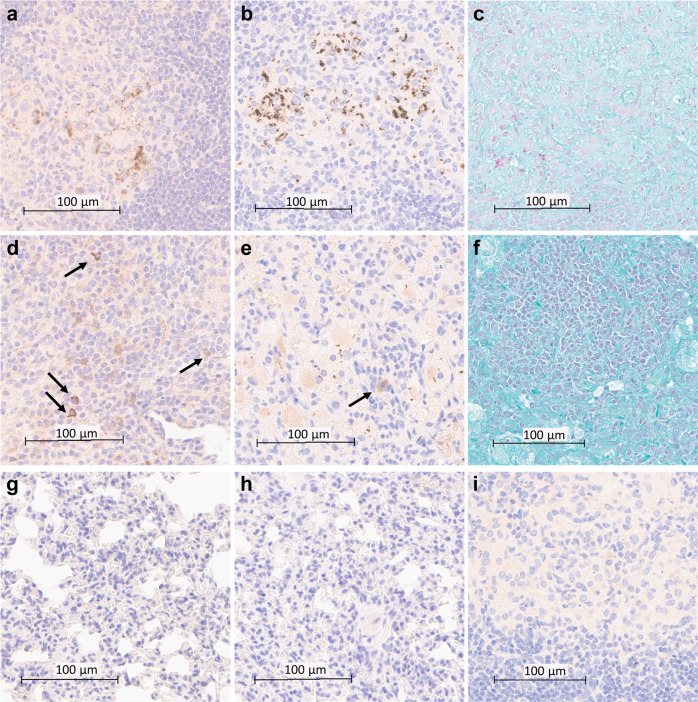
Fig. 8. T1AM09 and L1AM04 detect extra- and intracellular Mtb and LAM in lung tissues of Mtb-infected mice.
Histology and immunohistochemistry of Mtb (CDC1551) infected murine lung (scale bar 100 µm) showing intra- and extracellular staining of Mtb by a T1AM09; b L1AM04; and c staining for Acid-Fast Bacilli (AFB); d Staining of intracellular LAM in Mtb (CDC1551) infected lungs (arrows indicate LAM within macrophages) by T1AM09; e Staining of intracellular LAM and single bacilli in Mtb Erdman infected lungs (arrow indicates LAM within macrophages) by L1AM04; and f lack of positive AFB staining of Mtb (Erdman) in approximately the same Mtb infected lung section as shown in the other figure panels. Overall lack of staining of non-infected murine tissue (scale bar 100 µm) by g T1AM09; and h L1AM04. i Lack of staining of Mtb (CDC1551) infected lung tissue by isotype-matched control mAb to a flavivirus (scale bar 100 µm). All mAbs were tested at 2 µg/mL.

