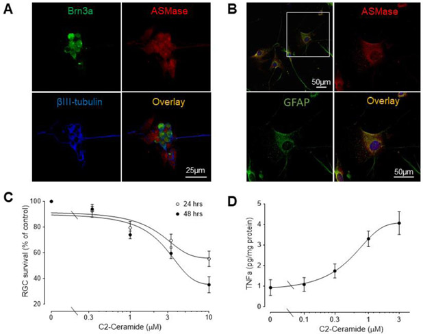
Figure 2. C2-ceramide decreases iPSC-RGC numbers and increases astrocyte TNF- α secretion.
(A) Localization of ASMase in human iPSC-derived RGCs. Immunofluorescence staining of Brn3a (green), ASMase (red), βIII-tubulin (blue) and overlay images. Scale bar, 25 μm. (B) Localization of ASMase in primary cultured human astrocytes. Immunofluorescence staining of GFAP (green), ASMase (red), and overlay images. Scale bar, 25 μm. (C) Survival assay of RGCs treated with C2-ceramide for 24 or 48 hours. Results were normalized to the cell numbers of vehicle-treated controls. Data are mean ± SEM, n≥4. (D) Effect of the C2-ceramide on TNF-α secretion in astrocytes. Cells were treated for 6 hours and media then collected and analyzed. Data are mean ± SEM; n ≥ 4.

