
Fig. 3.
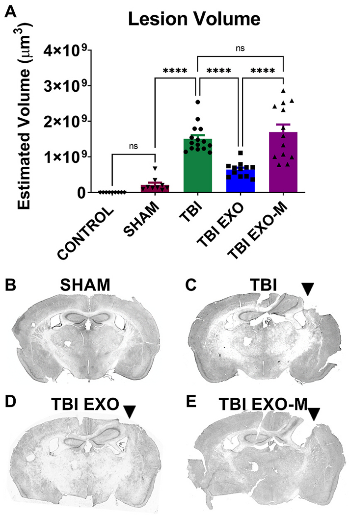
Exosomes reduce the impact lesion size following CCI.
Hematoxylin QS staining was utilized to quantify the impact area following TBI in the ipsilateral hemisphere. Using Cavalieri estimator to measure lesion volume treatment with exosomes (TBI+EXO) significantly reduced lesion volume compared to untreated TBI animals (p < 0.001 One way ANOVA F(4,52) = 36.99 followed by Tukey’s multiple comparisons) 6 weeks post TBI. However, treatment of exosomes depleted of MALAT1 (TBI+EXO-M) did not show rescue following injury when compared to untreated TBI animals. Tukeys post-hoc comparisons are indicated by asterisks on the graph ****p < 0.0001.