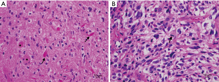
Figure 1.
H&E stain of patient’s tumor from 2006 (A) compared to 2020 (B). (A) Pilocytic astrocytoma with low cellularity and scattered piloid cells. Rosenthal fibers (arrows) and eosinophilic granular bodies (stars) are present. (B) Recurrent tumor showing increased cellularity and larger pleomorphic elements. Mitotic figures are present (arrow). H&E, hematoxylin and eosin.

