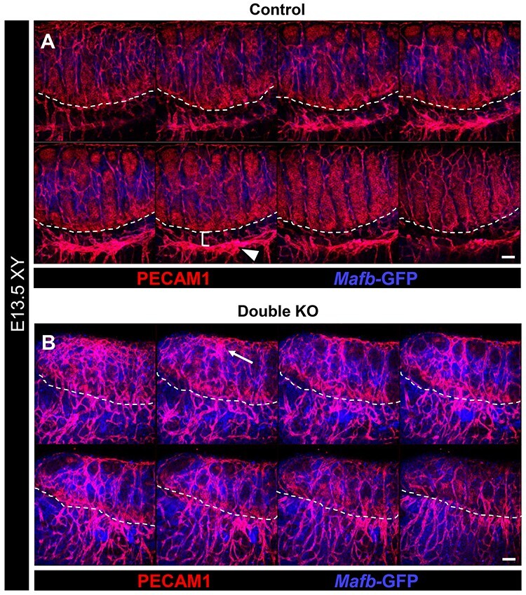
Figure 4.

Double KO testes exhibit disruptions in vascular patterning. Immunofluorescent images of E13.5 control (A) and double KO (B) fetal testes. Each panel shows eight consecutive confocal optical sections equally spaced through the entire gonad. Dashed lines indicate gonad-mesonephros boundary. Control testes (A) possess a fully developed gonad-mesonephric vascular plexus (arrowhead in A) with a well-defined avascular region between the vascular plexus and gonad (bracket in A). In contrast, double KO gonads (B) exhibit severely disrupted vascular development, with a hypervascularized coelomic surface (arrow) and aberrantly vascularized gonad-mesonephric border region. Scale bars, 100 μm.
