Figure 5.

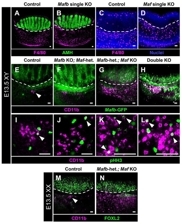
Maf mutant gonads display supernumerary CD11b-bright immune cells. Immunofluorescent images of E13.5 XY control (A, C, E, and I), Mafb single KO (B), Maf single KO (D), Mafb KO;Maf-heterozygous (F and J), Mafb-heterozygous; Maf KO (G and K), and double KO (H and L) gonads. Dashed lines mark gonad-mesonephros boundary. (A–D) F4/80+ differentiated macrophages are detected in similar numbers between control (A and C), Mafb single KO (B), and Maf single KO (D) fetal testes. However, in both Mafb-heterozygous; Maf KO (G) and double KO (H) testes, there is a dramatic increase in CD11b-bright cells relative to the small clusters of cells (arrowhead in E) found in control samples. (I–L) All genotypes exhibit mitotic (pHH3+) CD11b-bright cells (arrowheads in I–L). (M and N) Compared with E13.5 XX control ovaries (M), which contain a cluster of CD11b-bright cells along the gonad-mesonephros border (arrowhead in M), E13.5 XX Mafb-heterozygous; Maf KO fetal ovaries (N) possess supernumerary CD11b-bright cells throughout the mesonephros. Scale bars, 50 μm.
