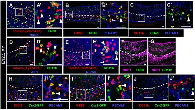
Figure 6.

CD11b-bright cells are fetal monocytes specifically localized near the gonad-mesonephric vascular plexus. Immunofluorescent images of E12.5 XY Vav1-Cre; Rosa-Tomato (A), wild-type C57BL/6 J (B, C, F, G), Lyz2-Cre; Rosa-Tomato (D), Csf1r-Cre; Rosa-Tomato (E), and Ccr2-GFP (H–J) gonads. A′–E′ and H′–J′ are higher-magnification images of the boxed regions in A–E and H–J. White dashed lines indicate gonad-mesonephros border throughout the figure. Labeling all hematopoietic cells with Tomato via Vav1-Cre (A) reveals both F4/80+ macrophages (arrow) and F4/80-negative round cells (arrowheads). (B, C) Staining with CD45 reveals F4/80-bright and CD11b-dim macrophages (B′ and C′, arrows), as well as F4/80-dim/negative, CD11b-bright round cells (B′ and C′, arrowheads). (D, E) Targeting myeloid cells with Lyz2-Cre (D) and monocyte/macrophages with Csf1r-Cre (E) reveals Tomato+ macrophages (D′ and E′, arrows; AIF1+ in D′) and CD11b-bright round cells (D′ and E′, arrowheads). (F, G) Whereas F4/80+ macrophages are associated with vasculature throughout the entire gonad-mesonephros complex (F), CD11b-bright cells are specifically localized near the gonad-mesonephric vascular plexus (G). NRP1 labels endothelial cells. (H–K) The monocyte marker Ccr2-GFP reveals GFP+/CD45+ cells near the gonad border (H′, arrowhead), which are occasionally F4/80+ (I′, arrowhead) and CD11b-bright (J′, arrowhead), but are mostly only GFP+. Scale bars, 50 μm.
