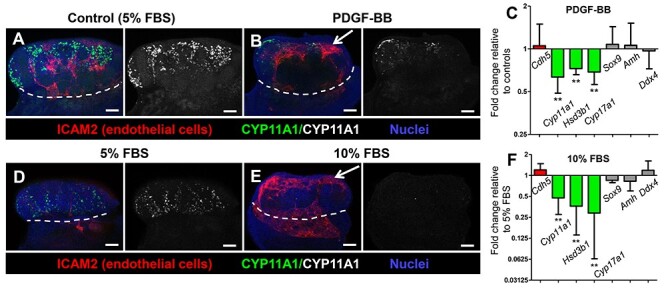
Figure 8.

Disrupted vascular patterning in the fetal testis is associated with reduced Leydig cell differentiation. Immunofluorescent (A, B, D, E) and qRT-PCR (C, F) analyses of 48-h ex vivo gonad culture of E12.5 CD-1 gonads, showing that disruptions in vascular patterning (arrows in B and E) caused by either PDGF-BB treatment (A–C) or increase in FBS concentration in the culture media (D–F) resulted in a decreased number of Leydig cells without effects on Sertoli or germ cells. White dashed lines indicate gonad-mesonephros border. Scale bars, 100 μm. All graph data are represented as mean ± SD. **, P < 0.01 (Student t-test).
