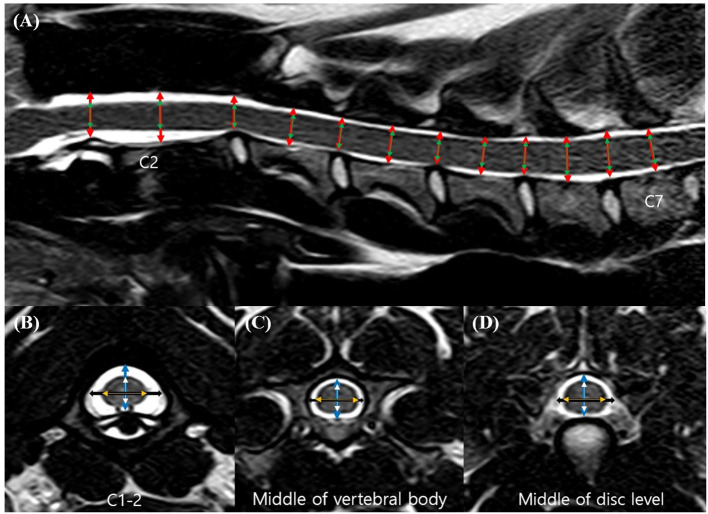
Figure 1.
Sagittal (A) and transverse (B–D) T2-weighted magnetic resonance (MR) images. Sagittal location of the measurements: the height of the spinal canal (red arrows) and the height of the spinal cord (green arrows). Transverse location of the measurements: the height of the spinal canal (blue arrows), the height of the spinal cord (white arrows), the width of the spinal canal (black arrows), and the width of the spinal cord (orange arrows).

