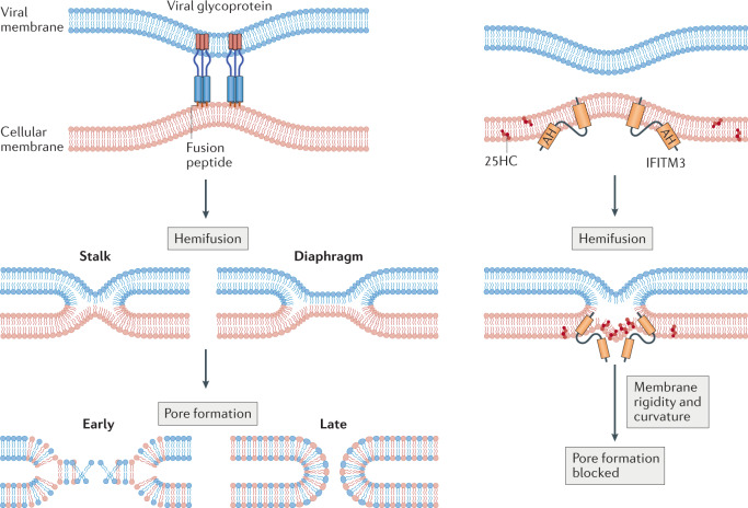
Fig. 2. Stages of virus–cell membrane fusion and the antifusion activities of IFITM3 and 25-hydroxycholesterol.
Close apposition of viral and cellular membranes is induced by viral envelope glycoproteins (shown at the upper left and not drawn thereafter), followed by the formation of a hemifusion stalk. Hemifusion is characterized by lipid mixing between the outer leaflets and alignment of the inner leaflets of each bilayer and may progress from a stalk to a diaphragm-like structure. Finally, further lipid mixing leads to partial opening of the fusion pore, and the pore is further dilated to complete membrane fusion. Interferon-induced transmembrane protein 3 (IFITM3) promotes hemifusion while inhibiting pore formation in a process that requires its amphipathic helix (AH) and protein oligomerization. IFITM3 promotes membrane rigidity and curvature in a manner that may disfavour formation of the fusion pore, and 25-hydroxycholesterol (25HC) produced by cholesterol 25-hydroxylase (CH25H) may function similarly.

