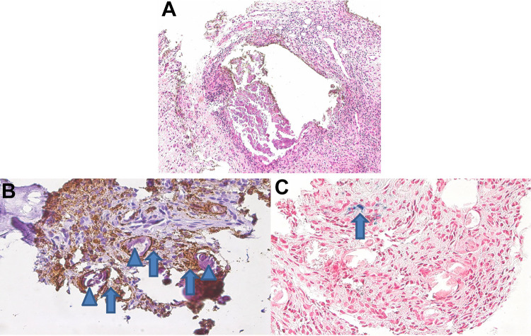
Figure 1.
(A) Bulky calcifications demarked by scarring fibrosis and marked inflammation (hematoxylin and eosin, 200×). (B) Calcified fragments (arrowheads) were surrounded by CD68-positive macrophages (arrows); CD68 immunostaining (brown), 400×. (C) Adjacent to the areas with inflammation, there were focal aggregates of hemosiderin (blue)-laden macrophages (arrow).

