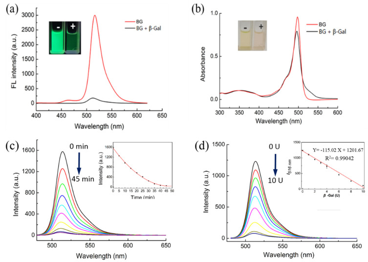
Figure 1.
Fluorescence and absorption changes of BOD-Gal to β-Gal (8 U) in DMSO/PBS solution (PBS / DMSO = 49:1 v:v, pH = 7.4). “-” indicated the absence of β-Gal, “+” indicated the presence of β-Gal. (a) Fluorescence changes, λex = 470 nm. (b) Absorption changes. (c) Time dependence of fluorescence spectra (0–45 min, λex = 470 nm). Inset: Curve of fluorescence intensity versus time. (d) Fluorescence changes of BOD-Gal to different concentration of β-Gal (0 U–12 U), λex = 470 nm. Inset: The relationship between I516 nm and the β-Gal concentration.

