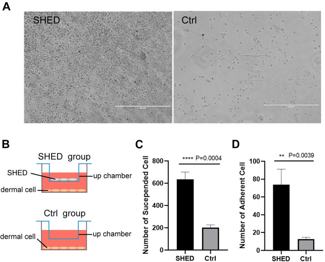
Figure 2.
SHED promote the growth of dermal cells in vitro (A) Freshly extracted C57BL/6 mouse dermal cells co-cultured with SHED for 3 days in a transwell chamber (left), and dermal cells without SHED co-culture (right); (B) Schematic diagram of co-cultivation of cells in Tranwell chamber. The gap between the upper and lower chambers is smaller than the cell diameter, and the two layers of cells can only communicate through signal molecules or cytokines; (C, D) Statistical results of the suspended and adherent cells in (A). Significance was calculated using t-test, P < 0.05 *, P < 0.01 **, P < 0.001 ***, P < 0.0001****.

