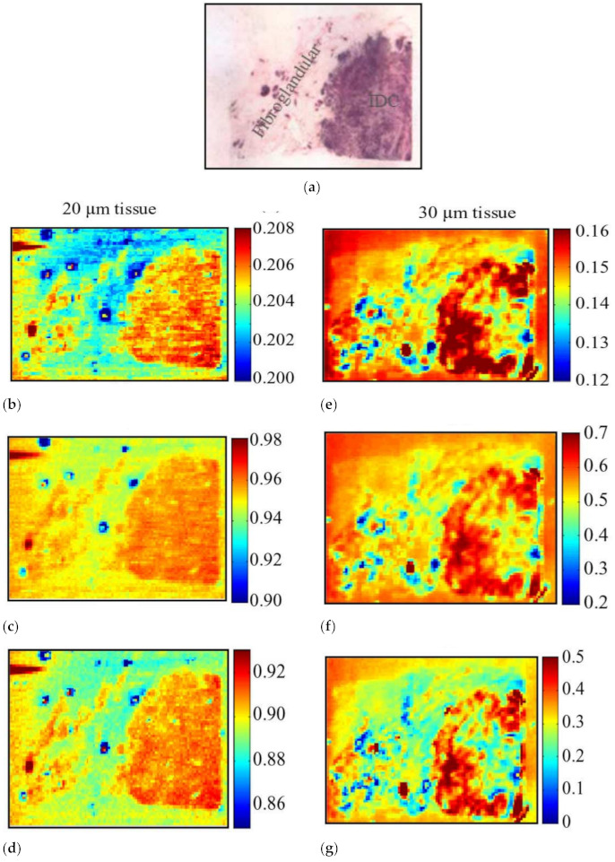
Figure 9.

THz image of sample 3. (a) Low-power H&E pathology image used for correlation. THz images for 20 µm: (b) time-domain image; (c) frequency-domain image at 1 THz; (d) frequency-domain image at 1.25 THz. THz images for 30 µm: (e) time-domain image; (f) frequency-domain image at 1 THz; (g) frequency-domain image at 1.25 THz [71].
