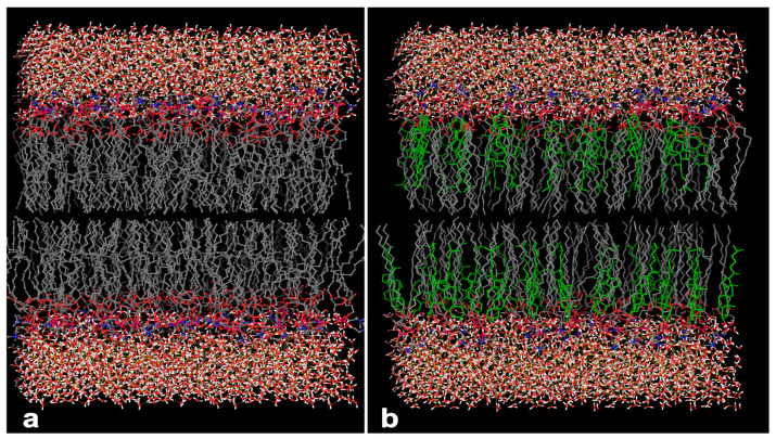
Figure 5.
Simulated 3D model of lipid bilayer. (a) Nano-phytosome prepared using lecithin; the polar site of phosphatidylcholine is represented with red color and the aggregation of white and red molecules at the top and bottom of the bilayer grid exhibit the water molecules the surround the lipid bilayer. (b) Phytosome prepared using lecithin conjugated with cholesterol; the green structure shows the arrangement of cholesterol within phosphatidylcholine.

