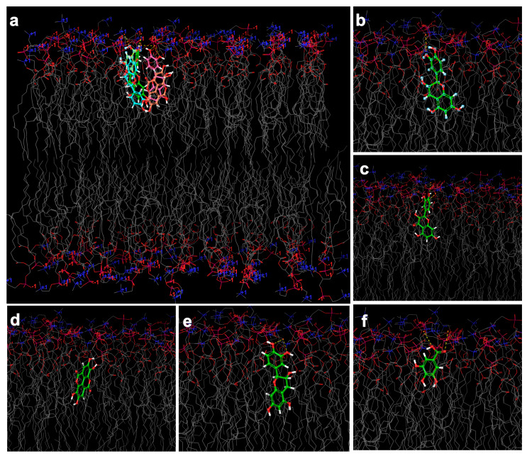
Figure 6.
Simulated 3D model of the nano-phytosome bilayer covered by thousands of water molecules (the red structure in the top and bottom). In real media, the bilayer appears as spherical, elliptical, or as a structure between both. The grid section of nano-phytosome bilayer is shown. The molecular docking images present the phenolic compound placed within the nano-phytosome bilayer structure with a number of hydrogen bindings; (a) overlaid of all compounds interaction with the bilayer, (b) catechin, and (c) epicatechin; both interacted through the 1,3-benzenediol site with the polar site of lipids with lower docking score and stronger hydrogen binding. (d) Ellagic acid, (e) quercetin, (f) gallic acid.

