
Figure 5.
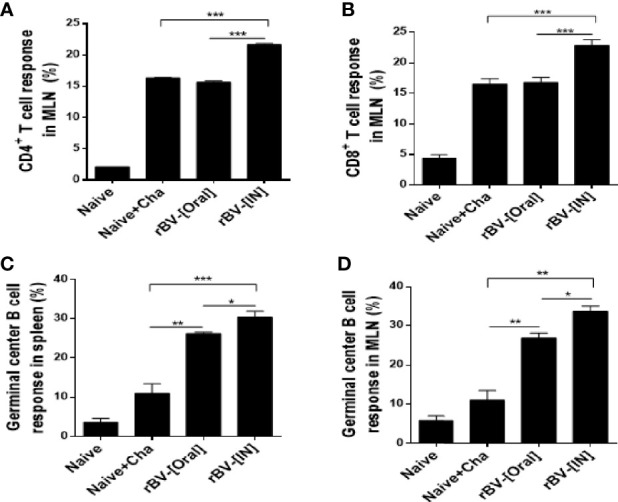
Detection of CD4+ T cell, CD8+ T cell, and GC B cell. Mice were sacrificed at 16 dpi for spleen and MLN acquisition. Single cell suspensions of splenocytes and MLN cells were prepared and analyzed using flow cytometry to confirm proliferation of CD4+ T cell (A), CD8+ T cell (B), and GC B cell (C) in MLN. Splenic GC B cell activation was also confirmed using flow cytometry (D). Data are presented as mean ± SD and asterisks denote statistical differences between groups. (*P < 0.05, **P < 0.01, ***P < 0.001).