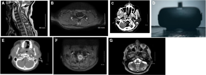
Figure 1.
MRI of the cervical spine, sagittal view (A) showing spinal canal stenosis and cord signal change, and axial view at C5–C6 (B). CT head shows hyperdense intracranial right vertebral artery (C) which was subsequently confirmed to be an arterial dissection on CT angiogram (white arrow indicating patent left vertebral artery, black arrow indicating occluded right artery) (E) and MRI dIssection protocol (arrows showing patent left vertebral artery and occluded right vertebral artery) (F). MRI brain showing matured left cerebellar infarct (G). Example of a currently available consumer virtual reality headset (photo by Christine Sandu, via Unsplash) (D).

