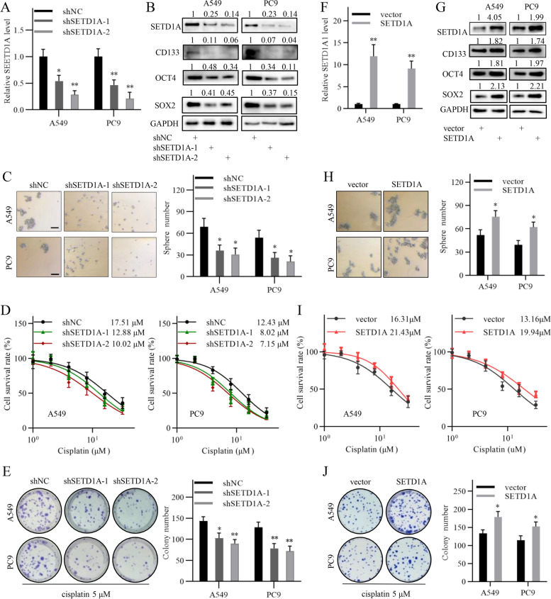
Fig. 2.
SETD1A regulates cancer stem cell property and cisplatin sensitivity in NSCLC cells. A, QRT-PCR analysis of SETD1A transcript expression in NSCLC cells transfected with shNC or shSETD1A lentivirus. B, SETD1A, SOX2, OCT4 and CD133 protein expression in NSCLC cells in the SETD1A knockdown and negative control groups was analyzed by western blotting. C, Sphere formation assay of the SETD1A knockdown and negative control NSCLC cells. Scale bar, 100 μm. D, Cisplatin sensitivity of the SETD1A knockdown and negative control NSCLC cells was analyzed by CCK-8 assay. The final concentrations of cisplatin were 1 μM, 2 μM, 4 μM, 8 μM, 16 μM and 32 μM. E, The growth of the SETD1A knockdown and negative control NSCLC cells exposed to cisplatin treatment was analyzed by colony formation assay. The final concentration of cisplatin was 5 μM. F, QRT-PCR analysis of SETD1A transcript expression in NSCLC cells transfected with the empty vector or SETD1A expression vector. G, SETD1A, SOX2, OCT4 and CD133 protein expression in the NSCLC cells transfected with empty vector or SETD1A expression vector was analyzed by western blotting. H, Sphere formation assay of NSCLC cells transfected with the empty vector or SETD1A expression vector. Scale bar, 100 μm. I, Cisplatin sensitivity of the NSCLC cells transfected with the empty vector or SETD1A expression vector was analyzed by CCK-8 assay. J, The growth of the NSCLC cells transfected with empty vector or SETD1A expression vector was analyzed by colony formation assay. The final concentration of cisplatin was 5 μM. Data are shown as means ± SD. *P < 0.05, **P < 0.01.

