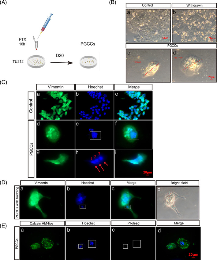
FIGURE 1.

PTX treatment can induce PGCC formation. (A) After PTX treatment for 16 hours and at 20 days posttreatment, TU212 was induced to form PGCCs. (B) (a) Normal TU212 without PTX treatment. (b) Cell morphology was observed after 16 hours of PTX treatment. (c,d) After 20 days, giant nuclei developed. Scale: 20 μm. (C) Cytoplasm was stained with vimentin (green) and nuclei were stained with Hoechst (blue). (a‐c) Control group. (d‐i) Multicore PGCCs; red arrow indicates nucleus. Scale: 20 μm. (D) (a‐d) Budding PGCCs, with DNA present in PGCC branches. Red arrow indicates nucleus. Scale: 20 μm. (E) PGCCs were alive. Images taken under 400× microscope
