
FIGURE 3.
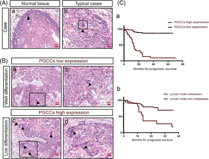
Number of PGCCs in tissue sections of patients with laryngeal carcinoma. (A) (a) HE staining of normal epithelial tissues. (b) HE staining of PGCCs, indicated with the black arrow. Scale: 20 μm. (B) (a and b) PGCCs in tissue sections from patients with well‐differentiated laryngeal carcinoma. (c and d) PGCCs in tissue sections of patients with lowly differentiated laryngeal carcinoma. (C) (a) Relationship between total survival time and PGCC count in patients with laryngeal cancer from 2013 to 2020. (b) Relationship between 3‐year survival rate and lymph node metastasis in patients with laryngeal cancer from 2013 to 2016, P < .05