FIG 1.
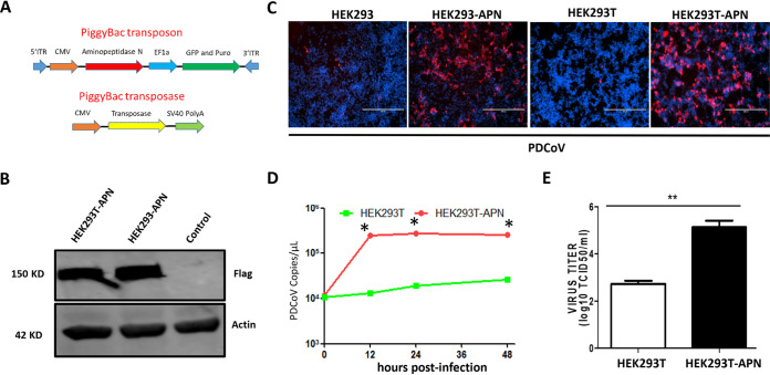
pAPN expression allows PDCoV replication in nonsusceptible HEK293 and HEK293T cells. (A) Scheme of the PiggyBac transposon system. ITR, internal transcribed spacer region; CMV, cytomegalovirus; SV40, simian virus 40. (B) HEK293-APN or HEK293T-APN cells were plated into six-well plates, and after 24 h, stable pAPN expression was confirmed by Western blotting using an anti-Flag antibody, with actin as the loading control. (C) HEK293, HEK293-APN, HEK293T, and HEK293T-APN cells were infected with PDCoV at an MOI of 0.1 in the presence of 200 ng/ml trypsin. PDCoV N protein was analyzed by immunofluorescence at 48 h postinfection (hpi). (D and E) Viral RNA in HEK293T and HEK293T-APN cells was collected at the indicated times (hours postinfection) and quantified by qPCR (D), and viral titers were quantified at 72 h (E). Each experiment was repeated at least three times. Error bars represent standard deviations (SD). *, P < 0.05; **, P < 0.01. TCID50, 50% tissue culture infective dose.

