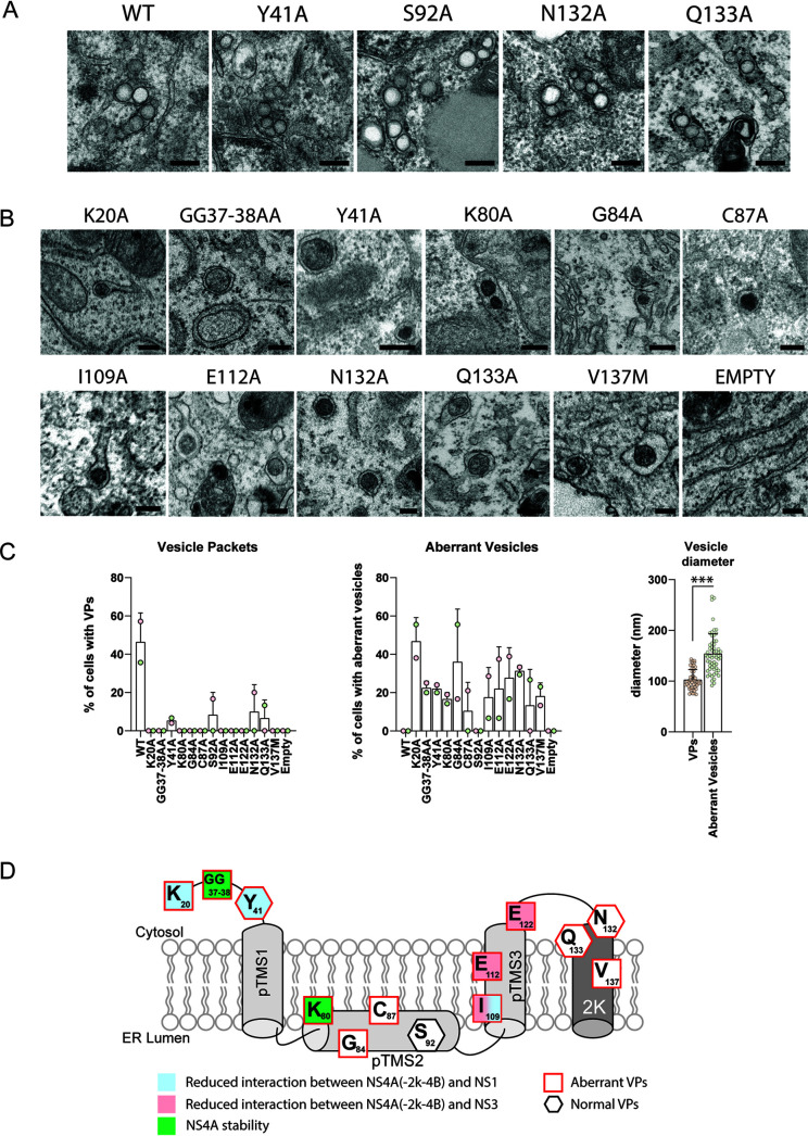
FIG 7.
Effect of NS4A mutations on the biogenesis of vesicle packets. (A and B) Electron microscopy analysis of Huh7/Lunet-T7 cells transfected with polyprotein constructs specified in Fig. 6A. Transfected cells were fixed and embedded into Epon resin, and thin sections were examined by transmission electron microscopy. For each sample, at least 15 cells were analyzed. Representative examples of vesicle packets or aberrant vesicles are shown in panels A and B, respectively. The experiment has been repeated twice. Scale bar, 200 nm. (C) Left and middle panels show the percentage of cells presenting VPs and aberrant vesicles, respectively. Each dot represents the mean of at least 15 cells analyzed. Two independent experiments were performed. The right panel shows the diameter of aberrant vesicles (nm) as found in cells transfected with the mutants specified in the middle panel. Each dot represents one vesicle. Means and SD are shown. ***, P < 0.001, as determined by two-tailed t test. (D) Graphical summary of the NS4A mutation analysis. Residues in NS4A and their contributions to interaction with NS1 and NS3, as well as their role in protein stability and VP formation, are displayed. Note that mutations affecting residues Y41, N132, and Q133 induced both regular VPs and VPs of aberrant morphology.

