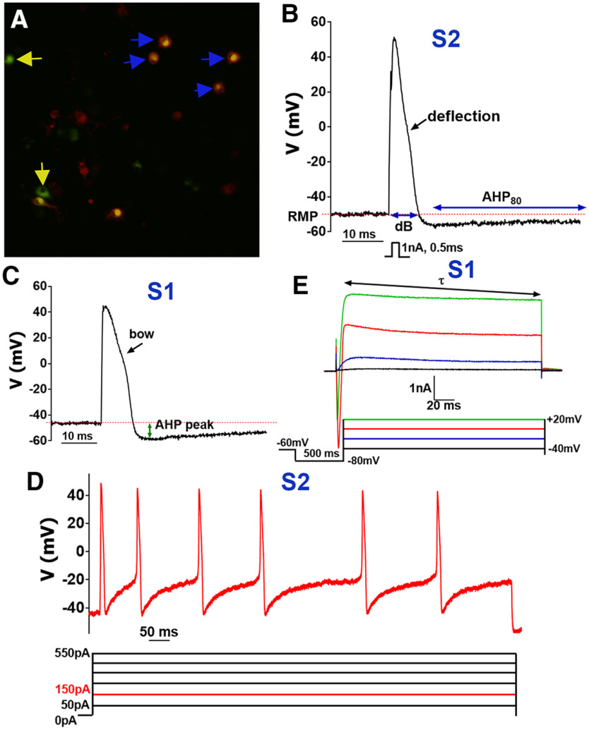
Figure 1.
Recordings from non-peptidergic IB4+ MM TG neurons. A, WGA-488+/IB4-555+ (marked with blue arrows), but not WGA-488+/IB4– (marked with yellow arrows) were selected for recording non-peptidergic TG neurons innervating MM. B, Stimulus waveform (1 nA, 0.5 ms) indicated below trace generated a single AP in a WGA+/IB4+ TG neuron belonging to the S2 group (Table 1). AP width is duration at base, dB. AHP80 is the time required for the AHP (measured in mV) to decay by 80% to a RMP level. Characteristic AP deflection is indicated by black arrow. C, AP from a WGA+/IB4+ TG neuron belonging to the group S1 (Table 1). Distinctive AP feature, bow, is indicated by black arrow. Distance from RMP to a lowest point of AP, AHP peak, is measured as indicated with the green arrow. D, Current-evoked AP train from a WGA+/IB4+ TG neuron belonging to the S2 group. Current waveforms are below trace and are applied by steps from 50 to 550 pA with 100-pA increment. Depictured AP train is evoked by a 150-pA step lasting 1 s. E, Currents were generated from a WGA+/IB4+ TG neurons belonging to the S1 group by the indicated waveforms found below traces. The decay constant τ was derived from standard single exponential fits between points indicated by arrows for the outward portion of the final current trace (+20 mV).

