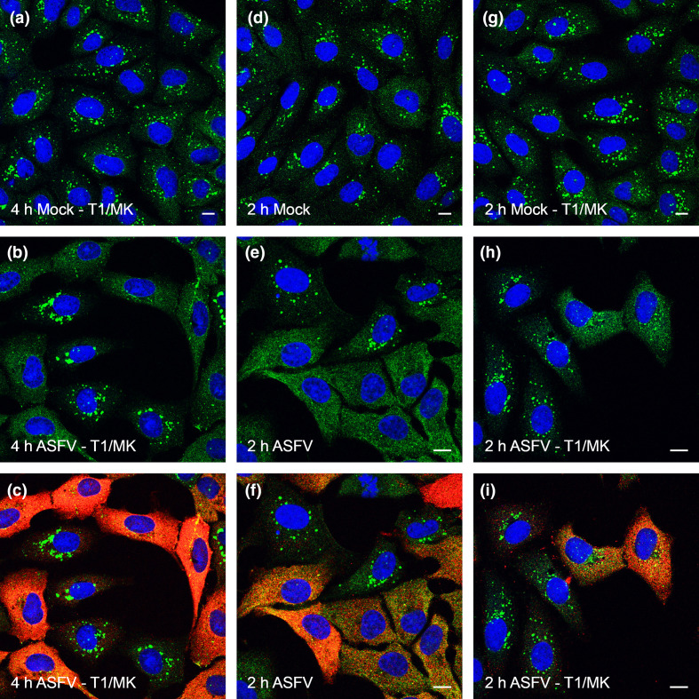
Fig. 5.
Biomodal inhibition of autophagosome formation by ASFV. Vero cells were incubated with mock inoculum (Panel a, d, g) or Ba71V (MOI 5) (Panels b, c, e, f, h, i) for 1 h. Inocula were removed and cells were incubated for a total of either 2 h (a–f) or 4 h (g–i) either in the presence (d–i) or absence (a–c) of 200 nM Torin1 and 5 µM MK-2206 (T1/MK). Cells were also starved by incubating with EBSS for the final 2 h of the incubation, i.e. throughout the 2 h incubation. Cells were then fixed and labelled with antibodies against LC3 (green), p30 (red) or with DAPI (blue). Panels c, f show the same infected cells as Panels B and E respectively but with the red channel removed to allow for clearer observation of LC3 staining. Scale bars represent 10 µm.

