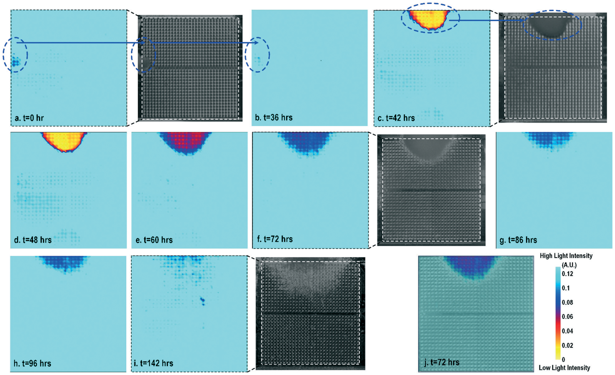
Fig. 2.

(a)–(i) Measured time-lapse optical opacity images of on-chip cultured fibroblasts for cell growth assays together with the corresponding reference stereo-microscope images. (j) The reference stereo-microscope image and the CMOS optical opacity image at t = 72 h are superimposed to show on-chip fibroblast location and matching. All optical opacity images use the same scale as that in (j).
