
Figure 5.
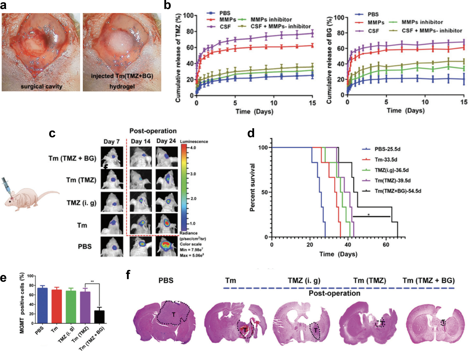
a) Photograph of the surgical cavity made by creating a cranial window through an incision in the midline to expose the brain for cutting the brain-tumor tissue (left) followed by injection of the therapeutic hydrogel (right) before sealing the cranial window. b) Cumulative release profiles of both TMZ (left) and BG (right) from Tm (TMZ + BG) hydrogel in PBS at 37 °C with gentle stirring under MMPs, MMPs + inhibitor, CSF and CSF + inhibitor conditions (data are presented as mean ± SEM, n = 4). c) Bioluminescence images of glioma-bearing mice 7, 14, and 24 days after post-operative treatment with PBS, Tm, TMZ (i. g.), Tm (TMZ), and Tm (TMZ + BG) hydrogel. d) Survival rate of the glioma-bearing mice after treatment. e) Percentage of MGMT positive cells as determined by immunohistochemistry staining of the tumor tissues dissected at day 26 after tumor implantation and treatment. f) H&E-stained brain sections where the dashed shape labeled T denotes tumor tissue.
Reproduced with permission. 2020, Royal Society of Chemistry.[67]