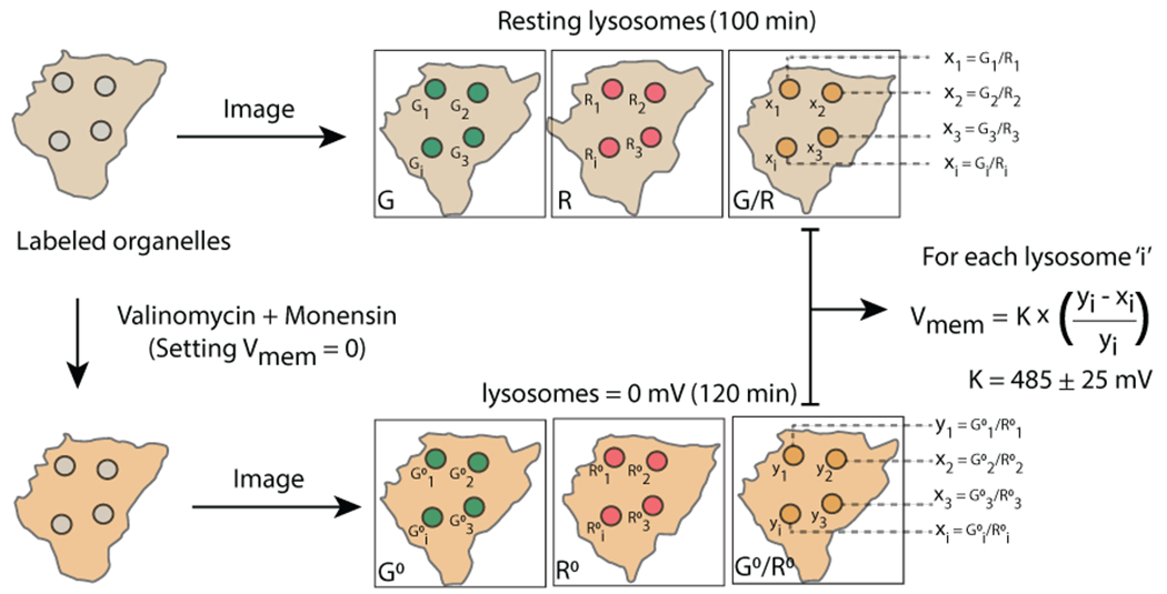
Extended Data Fig. 1.

Schematic of Lysosomal Vmem measurement, Imaging protocol of organelles labeled with VoltairIM. resting organelles are imaged in G and R channels, neutralized with valinomycin-monensin and second set of images are acquired in G and R channels. Resting membrane potential of the organelle is calculated by normalizing G/R values from untreated lysosomes (xi) to neutralized lysosomes (yi).
