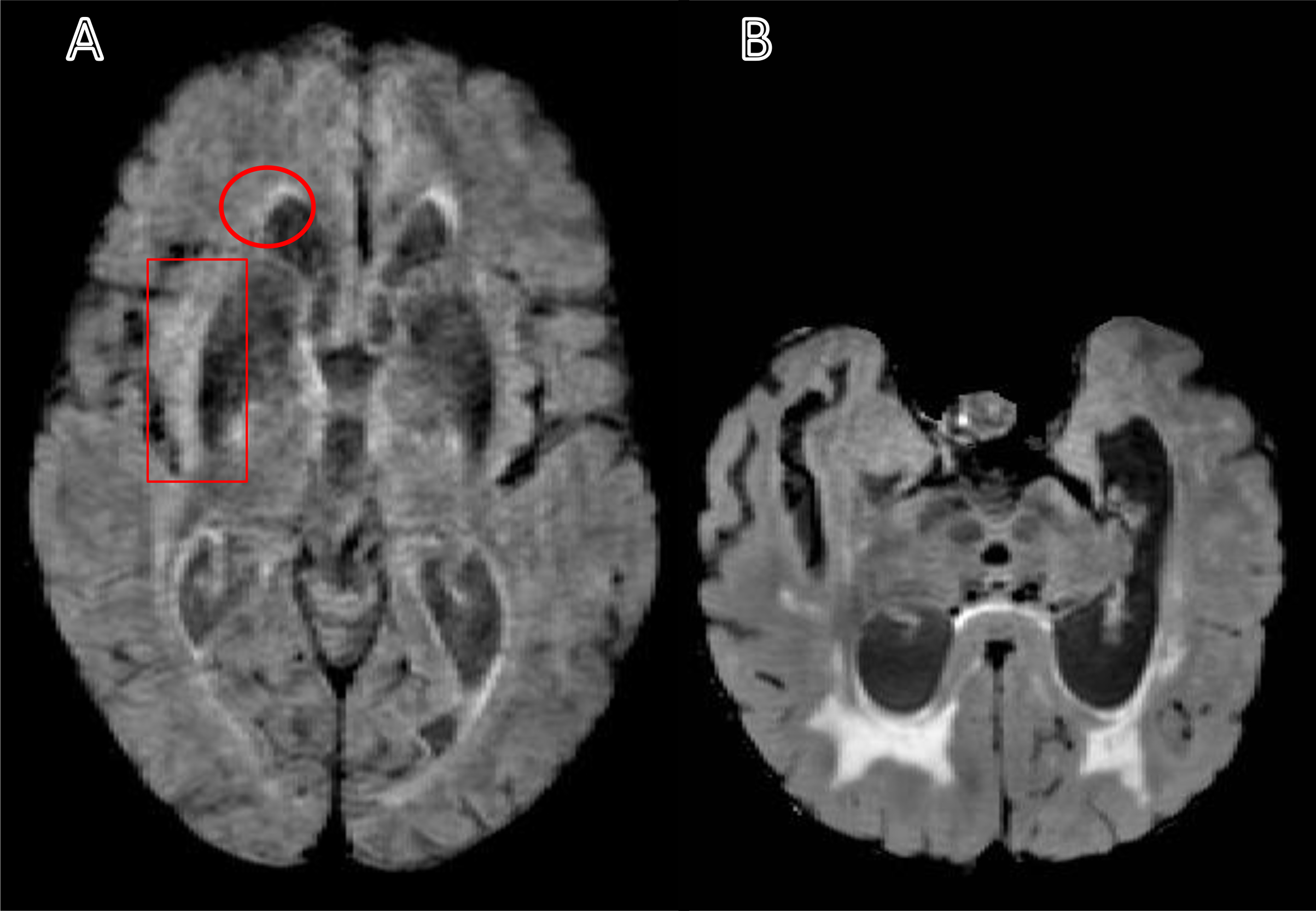
Fig. 1:

Sample of the excluded T2-weighted fluid-attenuated inversion recovery (FLAIR) images. A) A FLAIR image with a motion artifact leads to undefined ventricles and white matter hyperintensities edges (see the circle). Also, the motion artifact could lead to a variation in the intensity that may appear as a WMH and may cause an overestimation volume (see the rectangle). (B) A FLAIR image shows an irregular brain shape and ventricles that can cause a segmentation error.
Note: The motion artifact in panel A can be compared to panel B, and the asymmetry in panel B can be compared to panel A.
