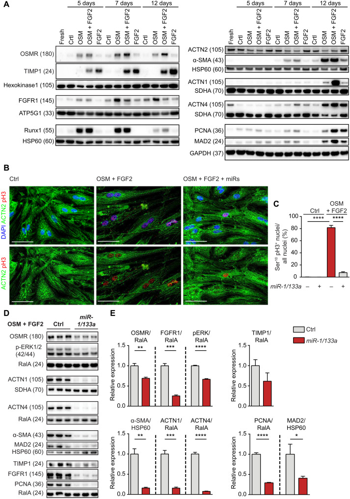
Fig. 2. Synergistic induction of cardiomyocyte dedifferentiation and cell cycle entry by OSMR and FGFR1 is inhibited by miR-1/133a.
(A) Western blot analysis of increased OSMR and FGFR1 expression and of markers related to cell cycle, fibrosis, and contractility after treatment of adult rat cardiomyocytes with OSM and FGF2. Different loading controls [HK1, HSP60, ATP5G1, SDHA, and GAPDH (glyceraldehyde-3-phosphate dehydrogenase)] were used for normalization to account for differences in molecular weights of proteins of interest on the respective membranes. (B and C) Immunofluorescence staining of adult rat cardiomyocytes showing increase of pH3 after stimulation with OSM and FGF2, which is inhibited by miR-1/133a (scale bars, 50 μm; n = 5 each group; unpaired two-tailed t test). ACTN2 staining was used to confirm the identity of cardiomyocytes. (D) Western blot analysis of OSMR and FGFR1 after stimulation of adult rat cardiomyocytes with OSM and FGF2 and miR-1/133a treatment. Expression of pERK and markers related to fibrosis, contractility, and cell cycle are shown as well. (E) Quantification of results shown in (D) (n = 3 each group; one-tailed unpaired t test). *P < 0.05, **P < 0.01, ***P < 0.001, and ****P < 0.0001.

