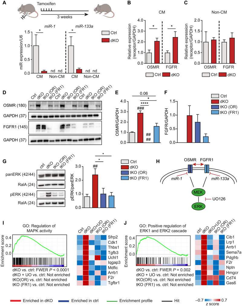
Fig. 3. Synergistic effects of OSMR and FGFR1 in mouse hearts after inactivation of miR-1/133a are mediated by MAPK-ERK.
(A) RT-qPCR analysis of miR-1/133a expression in adult dKO and control cardiomyocytes (n = 6, each) 3 weeks after tamoxifen administration. miR-1/133a is not detected (nd) in noncardiomyocytes (non-CM; ctrl, n = 5; dKO, n = 6; one-tailed unpaired t test). (B and C) RT-qPCR analysis of OSMR and FGFR1 expression in adult cardiomyocytes (B) (n = 4 each) and non-CM (C) (OSMR ctrl, n = 4; dKO, n = 3; FGFR1 ctrl, n = 5; dKO, n = 4; two-tailed unpaired t tests) of ctrl and dKO mice normalized to GAPDH 3 weeks after tamoxifen treatment. (D to F) Western blot analysis of OSMR and FGFR1 expression in ctrl, dKO, and tKO hearts (n = 3 each; one-tailed unpaired t test), normalized to GAPDH. (G) Western blot analysis of phosphorylated ERK in adult dKO compared to control and tKO hearts [ctrl, n = 5; dKO, n = 5; tKO (OR), n = 3; tKO (FR1), n = 3; one-tailed unpaired t test]. (H) Model of OSMR and FGFR1 repression by miR-1 and miR-133a. (I and J) Gene set enrichment analysis (GSEA) of microarray analysis data from adult mouse hearts. Genes related to MEK-ERK signaling are enriched in dKO versus ctrl hearts. Heatmaps of the 10 most up-regulated genes in dKO hearts derived from respective gene sets. Treatment with UO126 or additional deletion of OSMR (tKO OR) or FGFR1 (tKO FR1) [ctrl: n = 4; dKO, dKO + UO, tKO (OR), and tKO (FR1): n = 3] normalizes expression of up-regulated genes in dKO hearts. *P < 0.05 and ****P < 0.0001 compared to dKO; ##P < 0.01 and ###P < 0.001 compared to WT.

