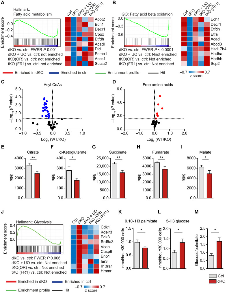
Fig. 6. Loss of miR-1/133a induces a metabolic shift in adult cardiomyocytes.
(A and B) GSEA of miR-1/133a knockout (dKO) versus WT hearts demonstrating down-regulation of genes related to fatty acid metabolism. Additional deletion of Osmr (tKO OR) or Fgfr1 (tKO FR1) in miR-1/133a dKO mice or UO126 treatment abolishes down-regulation. Heatmaps of the 10 most down-regulated genes in dKO hearts derived from the respective gene sets [ctrl: n = 4; dKO, dKO + UO, tKO (OR), tKO (FR1): n = 3]. (C to I) Metabolome analysis of dKO (n = 9) compared to control hearts (n = 12) showing significant decline of all detected acyl-CoA species (C) and increase of all detected free amino acids (D). Each dot in the volcano plots represents one specific metabolite. (E to I) Concentrations of metabolites demonstrate decreased TCA activity in miR-1/133a dKO hearts. (J) GSEA of microarray data from adult mouse hearts and heatmap of the 10 most up-regulated genes in dKO hearts derived from the respective gene set, showing enrichment of genes related to glycolysis in miR-1/133a (dKO) versus WT hearts. Additional knockout of Osmr (tKO OR) or Fgfr1 (tKO FR1) or UO126 treatment of dKO hearts abolished enrichment. (K to M) Assessment of palmitate and glucose catabolism, demonstrating decreased fatty acid oxidation (K) but increased glycolysis (L) and increased glycolysis/fatty acid oxidation ratios (M) in miR-1/133a dKO cardiomyocytes compared to ctrl (n = 8 to 9; one-tailed unpaired t test). *P < 0.05 and **P < 0.01.

