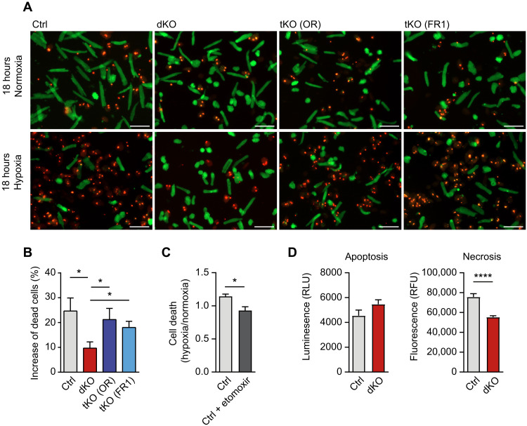
Fig. 7. Loss of miR-1/133a increases survival of adult cardiomyocytes under hypoxia.
(A and B) Cell viability assays of adult miR-1/133a dKO compared to ctrl cardiomyocytes under normoxia and hypoxia (18 hours; 1% O2). Increased survival of miR-1/133a dKO cardiomyocytes is prevented in tKO (OR) and tKO (FR1) mice [ctrl, n = 9; dKO, n = 7; tKO (OR), n = 5; tKO (FR1), n = 6; one-tailed unpaired t test]. Scale bars, 100 μm. (C) Cell viability assay of adult mouse cardiomyocytes without and with etomoxir (100 μM, 12 hours) under normoxia and hypoxia (18 hours, 1% O2). Etomoxir treatment increases the number of viable cardiomyocytes under hypoxia (n = 3 each; two-tailed unpaired t test). (D) Assays for apoptosis and necrosis using isolated adult mouse cardiomyocytes reveals no significant difference in apoptosis, but reduced necrosis of miR-1/33a dKO compared to control cardiomyocytes (n = 2 animals each, 4 technical replicates each). RLU, relative luminescence units; RFU, relative fluorescence units.

