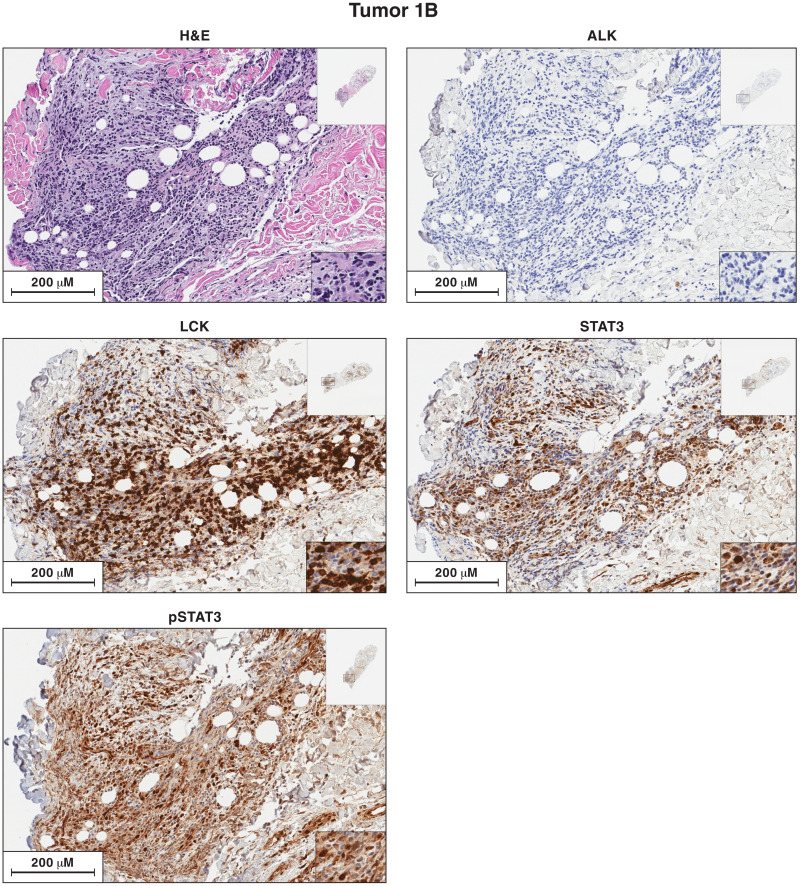
Fig. 3. Antibody staining of sections of sample 1B for the expression of ALK, LCK, STAT3, and pSTAT3.
Each image is labeled to show what the section was stained for. The procedures used to antibody stain the sections of the lymphoma samples are given in Materials and Methods. Sections from each of the lymphoma samples were H&E stained. Additional sections from each lymphoma were reacted separately with antibodies to ALK, LCK, STAT3, and pSTAT3. The images in the figures show a portion of the section. For each stained section from a particular lymphoma, the same section was chosen for the figure. The inset at the top right shows the whole section, and the inset at the bottom right shows a small part of the image at higher magnification. The images can be expanded to show more detail.

