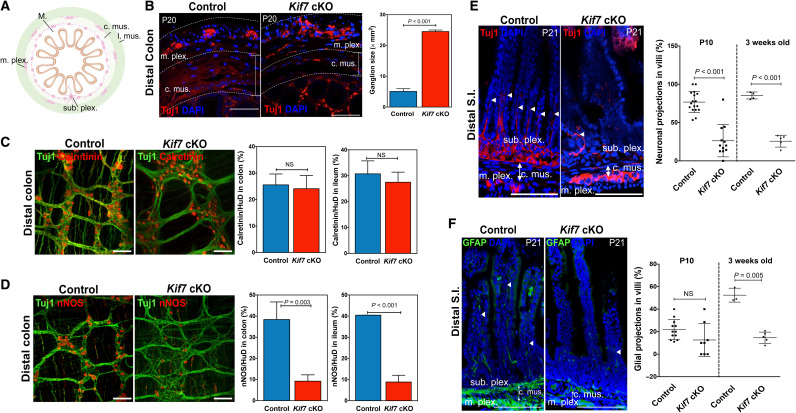
Fig. 2. Ablation of Kif7 interferes with the formation of myenteric and submucosal neurons and gut colonization.
(A) Schematic of the organization of the intestine. (B) Transverse sections of P20 control and Kif7 cKO colons. Myenteric ganglia were marked using anti-Tuj1 antibody; the size of the ganglia was measured and is shown in the bar chart. (C) The excitatory and inhibitory neurons in the myenteric plexuses of the distal colons of control and Kif7 cKO mutants (P21) were detected using antibodies against (C) calretinin and (D) nNOS, respectively. The numbers of excitatory (calretinin+) and inhibitory (nNOS+) neurons as a percentage of the total number of neurons (HuD+) were determined and are shown in the bar charts. Immunohistochemistry analyses for the detection of submucosal plexuses and (E) neuronal (Tuj1+) and (F) glial (GFAP +) processes in villi with the transverse sections of the distal small intestine of control and Kif7 cKO (P21). The neuronal and glial processes are marked with arrowheads. The percentages of villi with neuronal and glial processes were measured at P10 and P21 and are shown in the scatterplots. The error bars indicate ± SEM of the values obtained from at least six samples. c. mus., circular muscle; l. mus., longitudinal muscle; m. plex., myenteric plexus; sub. plex., submucosal plexus.; M., mucosa. Scale bars, 100 μm. DAPI, 4′,6-diamidino-2-phenylindole. NS, not significant.

