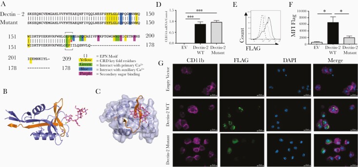
Figure 1.
Dectin-2 mutation results in minimal protein expression. A, Partial amino acid sequence of wild-type (WT) and mutant (507delC) Dectin-2 with key residues and EPN mannan-binding motif highlighted. B, The fold of WT Dectin-2 5VYB (cartoon) in complex with mannan (stick model). Blue = present in the mutant protein, orange = absent in the mutant protein. Two Ca2+ atoms (green spheres) and an Na+ atom (purple) are also displayed. C, Predicted surface model of Dectin-2 covering the mutant structural elements only. The final β-strand at the core of the structure is missing in the mutant, resulting in a collapse of the motif, corruption of ligand interface, and inability to bind mannan. D–G, Bone marrow–derived macrophages (BMDMs) from Dectin-1–Dectin-2 knockout (KO) mice were infected with constructs expressing FLAG-tagged Dectin-2 WT, mutant, or empty vector (EV) and harvested 72 hours later. D, RNA was isolated, complementary DNA was prepared, and CLEC6A messenger RNA (mRNA) transcript was detected by reverse-transcription quantitative polymerase chain reaction. mRNA levels were normalized to HPRT1. Graph displays mean ± standard error of the mean (SEM) from 3 independent experiments. One-way analysis of variance (ANOVA) with Tukey posttest on transformed data. E and F, Cells were permeabilized, stained with anti-FLAG, and analyzed by flow cytometry. E, Dashed black line = empty vector; solid black line = Dectin-2; solid gray line = Dectin-2 mutant. Histogram representative of 3 independent experiments. F, Graph displays mean ± SEM of mean fluorescence intensity from 3 independent experiments. One-way ANOVA with Tukey posttest. G, BMDMs were stained with anti-CD11b (magenta) and anti-FLAG (green); nuclei were stained with 4’,6-diamidino-2-phenylindole (blue). Images are representative of 2 independent experiments. *P < .05; ***P < .001. Abbreviations: DAPI, 4’,6-diamidino-2-phenylindole; EV, empty vector; MFI, mean fluorescence intensity; mRNA, messenger RNA; WT, wild-type.

