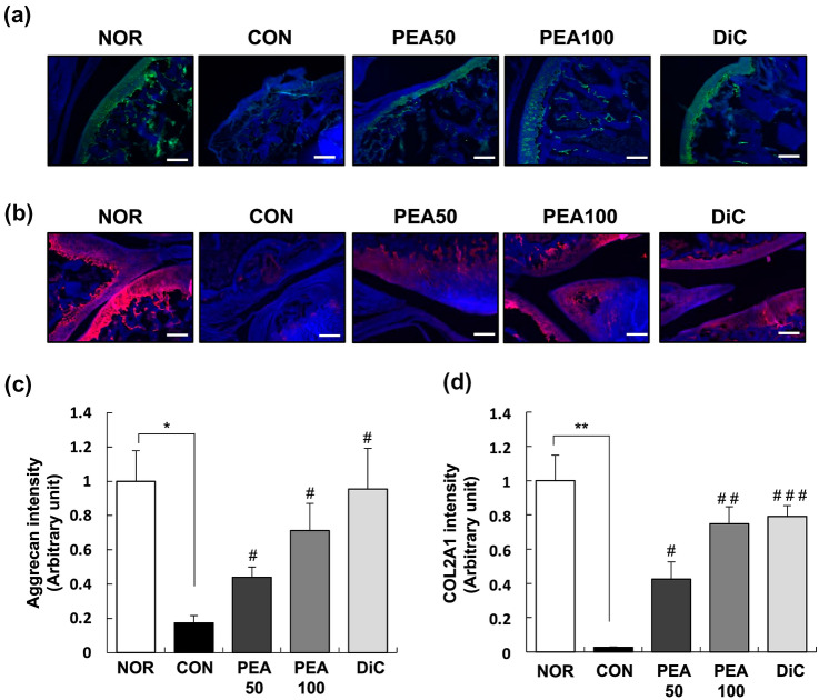
Fig. 3.
Effect of PEA on the expression of aggrecan and COL2A1 in the articular cartilage of MIA-induced OA rats. MIA-induced OA rats were administered either PEA (50 or 100 mg/kg BW/day) or diclofenac (6 mg/kg BW/day) for 4 weeks. Articular cartilage was stained with aggrecan (a) and COL2A1 (b) antibodies. Representative IF staining images are shown. Scale bar, 50 μm. (c), (d) Staining intensity of the indicated proteins was quantified. Each bar represents mean ± SEM (n = 5). *P < 0.01, **P < 0.05, and ***P < 0.001 significantly different from the NOR group. #P < 0.01, ##P < 0.05, and ###P < 0.001 significantly different from the CON group. COL2A1 collagen type II alpha 1, PEA palmitoylethanolamide, IF immunofluorescence, OA osteoarthritis, MIA monosodium iodoacetate, BW body weight, NOR normal control group (injected with saline + treated with phosphate-buffered saline (PBS)), CON control group (injected with MIA + treated with PBS), PEA50 or PEA100 50 or 100 mg/kg body weight (BW)/day PEA-treated group (injected with MIA + treated with 50 or 100 mg of PEA/kg BW/day), DiC positive control group (injected with MIA + treated with 6 mg of diclofenac/kg BW/day)

