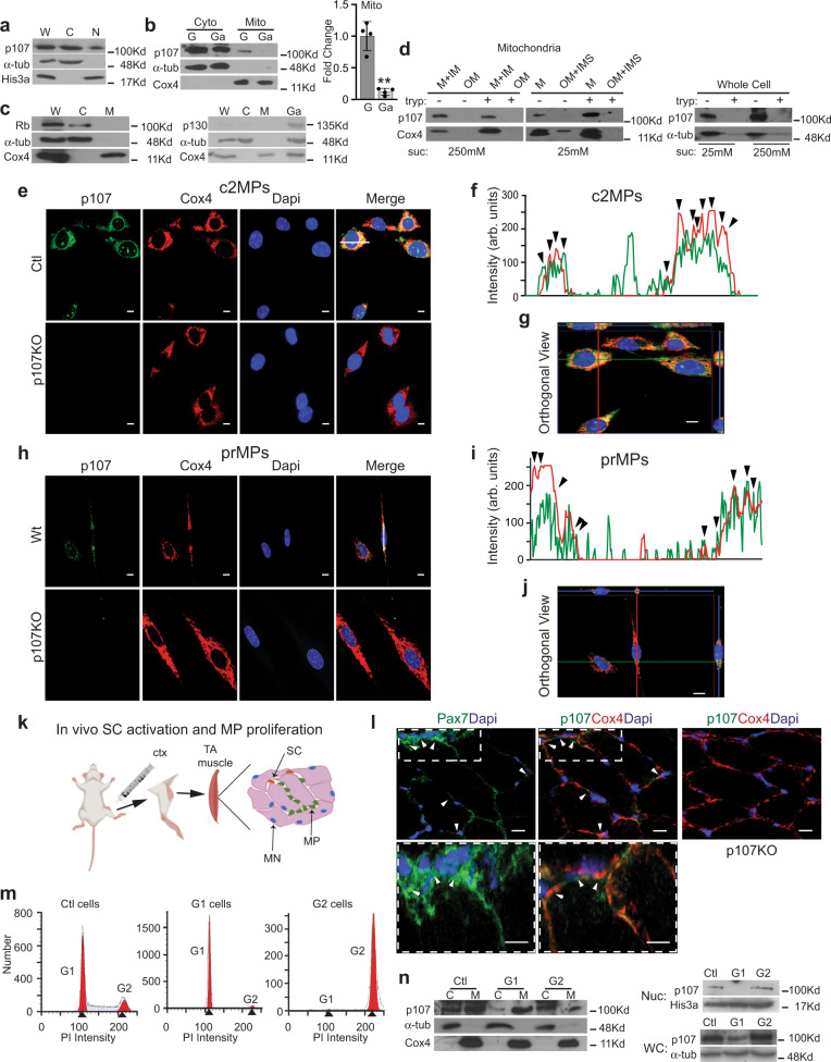
Fig. 1. p107 is localized in the mitochondria of proliferating myogenic progenitors.
a Representative Western blot of whole cell (W), cytoplasmic (C), and nuclear (N) fractions for p107, cytoplasm loading control α-tubulin (α-tub) and nucleus loading control, His3a, during proliferation. b Representative Western blot and graphical representation of cytoplasmic (Cyto) and mitochondrial (Mito) c2MP fractions for p107, α-tub and mitochondria loading control, Cox4, during proliferation (G) and growth arrest (Ga). Graphical data are presented as mean values ± SD (n = 4 biologically independent samples). Two-tailed unpaired Student’s t test **p = 0.00343. c Representative Western blot of proliferating whole cell (W) cytoplasmic (C), mitochondrial (M) and growth arrested (Ga) cells for Rb, p130, α-tub and Cox4. d Representative Western blot of c2MP whole cell and mitochondrial fractions including outer membrane (OM), inner membrane (IM), soluble inner membrane (IMS) and matrix (M) that were isolated in 250 mM or 25 mM sucrose buffer, treated and untreated with trypsin. Confocal immunofluorescence microscopy for p107, Cox4, Dapi and Merge of proliferating e Control (Ctl) and genetically deleted p107 (p107KO) c2MPs and h wild type (Wt) and p107KO primary (pr) MPs (scale bar 10 μm). f, i A line was drawn through a representative cell to indicate relative intensity of RGB signals with the arrowheads pointing to areas of concurrent intensities g, j an orthogonal projection was generated by a Z-stack (100 nm interval) image set using the ZEN program (Zeiss) in the XY, XZ, and YZ planes (scale bar 10 μm). k Schematic for inducing in vivo satellite cell (SC) activation and MP proliferation in the tibialis anterior (TA) muscle with cardiotoxin (ctx) injury (MN is myonuclei). l Confocal immunofluorescence microscopy merged image of wild type (Wt) TA muscle section 2 days post ctx injury for Pax7 and Dapi and p107, Cox4 and Dapi and for p107KO TA muscle section for p107, Cox4 and Dapi (scale bar 20μm). Arrows denote Pax7 +p107+Cox4+ cells. m Cell cycle histograms using flow cytometry for cell number versus propidium iodide (PI) intensity for c2MPs that are proliferating (Ctl), predominately in G1 and G2 phase of the cell cycle. n Representative Western blot of cytoplasmic (C), mitochondrial (M), nuclear (Nuc) and whole-cell lysates (WC) for p107, α-tub, Cox4 and His3a from Ctl, G1 and G2 cells. Source data are provided as a Source Data file.

