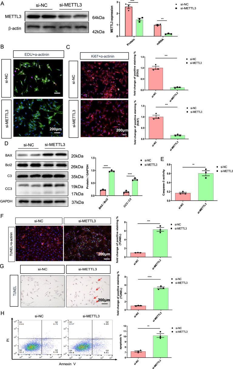
Fig. 3. METLL3 silencing mimics the effects of hypoxia on decreasing proliferative capacity and increasing apoptosis in NRCMs.
A Protein and mRNA expression of METTL3 in the NRCMs from indicated groups. B Representative immunofluorescence images of NRCMs labeled with EDU or α-actinin (EDU, red; α-actinin, green; DAPI, blue. Scale bars, 200 μm). C Representative immunofluorescence images of NRCMs labeled with Ki67 or α-actinin (α-actinin, red; Ki67, green; DAPI, blue. Scale bars, 200 μm). D Protein level of BAX, Bcl2, C3, and CC3 in the NRCMs. E Caspase 3 activities of NRCMs from indicated groups. F, G Representative immunofluorescence (F) and immunohistochemical (G) staining images of NRCMs labeled with TUNEL (α-actinin, red; TUNEL, green; DAPI, blue. Scale bars, 200 μm), and the corresponding quantitative analysis. H The results of flow cytometry on apoptosis in NRCMs. N = 3. The data were presented as the mean ± SD.

