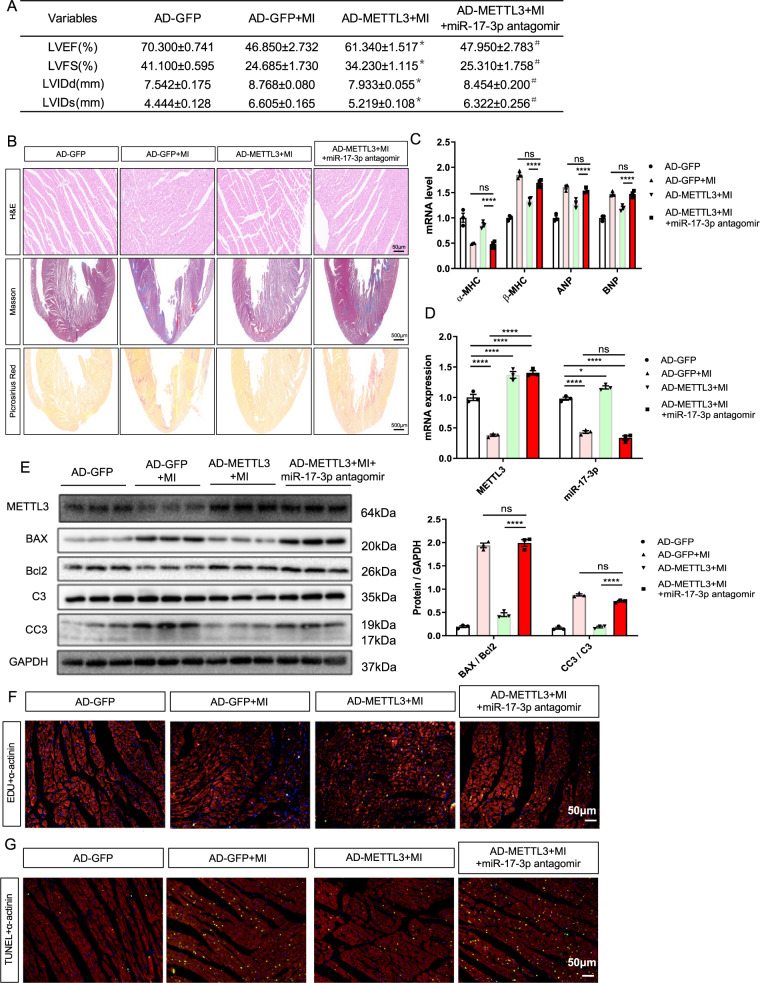
Fig. 6. miR-17-3p antagomir blocked the protective effects of METTL3 in myocardial infarction (MI)-induced rats.
A Echocardiographic data of left ventricular function, n = 5. B Representative images of non-infarcted left ventricular myocardium sections stained with hematoxylin-eosin (HE; Scale bar, 500 μm), Masson and picrosirius red (Scale bar, 50 μm). C mRNA expression of α-MHC, β-MHC, ANP, and BNP in rats from indicated groups, n = 5. D mRNA expression of METTL3 and miR-17-3p in non-infarcted left ventricular myocardium from different groups, n = 3. E Protein expression of METTL3, BAX, Bcl2, C3, and CC3 in non-infarcted left ventricular myocardium from indicated groups, n = 3. F, G Representative immunofluorescence images of non-infarcted left ventricular myocardium sections labeled with EDU (F) or TUNEL (G) (α-actinin, green; EDU or TUNEL, red; DAPI, blue. Scale bars, 50 μm). The results are expressed as the mean ± SD.

