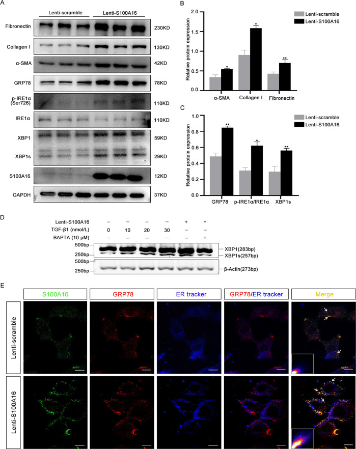
Fig. 4. S100A16 participates in endoplasmic reticulum stress in HK-2 cells through IRE1α/XBP1 pathway.
A Expressions of the renal fibrosis-related genes fibronectin, collagen I, and α-SMA, and the ER stress-related genes GRP78, p-IRE1α, XBP1, and XBP1s in S100A16-overexpressing and Lenti-scramble HK-2 cells. B Quantitation of fibronectin, collagen I, and α-SMA protein expression in A, normalized to GAPDH expression. C Quantitation of GRP78, XBP1s, and p-IRE1α/IRE1α protein expression in a, normalized to GAPDH expression. Bars indicate the means ± SE from three independent experiments. *P < 0.05, **P < 0.01, ***P < 0.001. D RT-PCR analysis of mRNA levels of spliced XBP1 in SA100A16-overexpressing HK-2 cells treated with TGF-β1 (0, 10, 20, and 30 nM) and/or with BAPTA-AM. E Immunofluorescence staining showing cellular colocalization of S100A16 and GRP78. S100A16 in green, GRP78 in red, and ER tracker in blue. Scale bar = 20 µm. The lower corner in the merged panel shows the pixels indicating colocalization of GRP78 and S100A16 in S100A16-overexpressing and Lenti-scramble HK-2 cells. The GRP78 and S100A16 colocalization pixels were identified by the Colocalization Finder plugin in ImageJ. Scale bar = 20 μm.

