Figure 3.
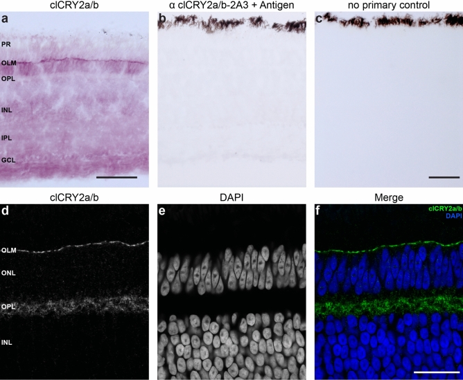
Localisation of clCRY2a/b in the pigeon retina. (a–f) Immunohistochemistry using the monoclonal clCRY2a/b antibody on adult pigeon retina harvested at midday (n = 3 birds). (a) Permanent staining reveals clCRY2a/b is localised throughout the retina and is enriched in the outer limiting membrane (OLM). (b) No staining is seen when 2A3 α clCRY2a/b is pre-incubated with the antigen. (c) Pigeon retinal sections treated with no primary clCRY2a/b antibody do not show any signal. (d–f) Immunofluorescence shows clCRY2a/b is enriched in the OLM and the outer plexiform layer (OPL). PR: photoreceptors, OLM: outer limiting membrane, ONL: outer nuclear layer, OPL: outer plexiform layer, INL: inner nuclear layer, IPL: inner plexiform layer, GCL: ganglion cell layer. Scale bars show 50 μm.

