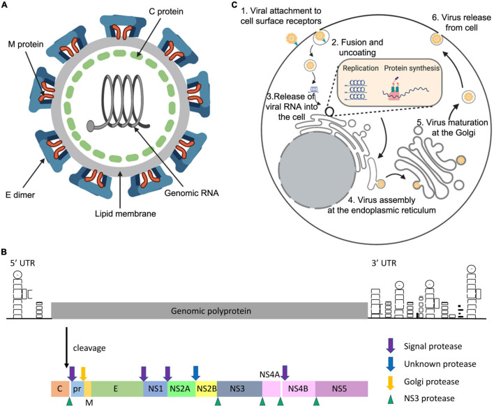
FIGURE 1.
General features of the ZIKV. The ZIKV virion (A) is an enveloped, spherical virus about 50 nm in diameter. The surface proteins envelope (E) and membrane (M) are arranged in an icosahedral structure that encases the nucleocapsid (C) protein bound to the viral RNA (adapted from viralzone.com and created with Biorender.com). The genome (B) is positive sense single-stranded RNA consisting of a single open reading frame, and 5′ and 3′ untranslated regions. The single synthesised polyprotein is post-translationally cleaved by host proteases (signal and Golgi peptidases) and the viral NS3 protease. This forms the three structural proteins: C, pre-membrane (prM), and E. Additionally, there are seven non-structural proteins which are necessary for genome replication and the modification of host cellular functions (adapted from Pierson and Diamond, 2018). (C) ZIKV enters the host cell using receptor mediated endocytosis via the receptors DC-SIGN, Tyrosine 3, TIM-1 or ACL. ZIKV is then translocated via endosome vesicles to the ER, and viral RNA is released. Translation occurs and the resulting polyprotein is subsequently cleaved using host and viral proteases. ZIKV remodels the ER and uses viral replication factories to carry out genome replication. Immature virions then assemble on the ER derived membrane, undergo maturation at the Golgi, and are released from the cell using the secretory pathway (adapted from Gerold et al., 2017, created with Biorender.com).

